Cytochrome C oxidase
Cytochrome c oxidase (abbreviated CCO, also known as complex IV) is the final and, from the perspective of red light therapy, the most important enzyme of the electron transport chain in mitochondria. It is a transmembrane protein complex that accepts electrons from cytochrome c, transfers them to molecular oxygen, and produces metabolic water. At the same time, it pumps protons and contributes to ATP production. And it is precisely CCO that is a chromophore, a molecule that absorbs red and near-infrared light. The entire science of photobiomodulation rests on this fact.
- What cytochrome c oxidase is and where it is located
- Which metal centers it contains and why they absorb light
- How CCO functions in the electron transport chain and why it is the final step to water production
- Why CCO is the key to photobiomodulation
- Which wavelengths CCO absorbs (the action spectrum according to Karu)
- How nitric oxide blocks CCO and how red light releases it
- What is cytochrome c oxidase and where is it found?
- What is the structure of CCO and why does it absorb light?
- What function does CCO serve in the electron transport chain?
- Why is CCO a chromophore for red and infrared light?
- Which wavelengths does CCO absorb?
- How does nitric oxide block CCO and how does light release it?
- What happens in the cell after light is absorbed by CCO?
- Why does CCO matter for your health?

What is cytochrome c oxidase and where is it found?
Cytochrome c oxidase is a large transmembrane enzyme embedded in the inner membrane of mitochondria. It is the fourth and final complex of the electron transport (respiratory) chain. In mammals, it consists of 13 subunits, of which the three largest (I, II, and III) are encoded by mitochondrial DNA and the remaining ten by the nuclear genome.
Richter and Ludwig (2003) described CCO as the terminal member of the electron transport chains of both mitochondria and many bacteria, providing an efficient mechanism for reducing oxygen while simultaneously pumping protons (Richter & Ludwig, 2003).
CCO is present in every cell that contains mitochondria, meaning virtually all cells in your body (with the exception of red blood cells, which lack mitochondria). It is found in muscle cells, neurons, keratinocytes of the skin, cells of the heart, liver, and immune system. Everywhere cells need energy.
What is the structure of CCO and why does it absorb light?
CCO contains four redox-active metal centers that are crucial for both its function and its ability to absorb light:
- CuA (copper center A) – a binuclear copper center in subunit II. This is the first site where electrons arrive from cytochrome c. CuA absorbs light primarily in the NIR range (760 to 850 nm).
- Heme a – a heme prosthetic group in subunit I. It transfers electrons from CuA to the binuclear center. It absorbs light in the red range (620 to 680 nm).
- Heme a3 – the second heme in subunit I, part of the binuclear center. This is where oxygen binds and is reduced to water.
- CuB (copper center B) – cooperates with heme a3 in the binuclear center to reduce oxygen. It absorbs in the NIR region.
Ishigami et al. (2023) described in detail in their study on the structural basis of CCO's functional properties how these four centers cooperate in electron transfer and oxygen reduction (Ishigami et al., 2023).
It is precisely these metal centers containing copper and iron that make CCO a chromophore, a molecule capable of absorbing photons. Red and infrared light has sufficient energy to interact with the d-orbitals of the metal ions in these centers, thereby changing the redox state of the enzyme and influencing its activity.
What function does CCO serve in the electron transport chain?
CCO is the final "station" of the electron transport chain. Its roles are:
- Accepts electrons from cytochrome c – a small soluble protein that carries electrons from complex III
- Reduces molecular oxygen to water – four electrons and four protons combine with one molecule of O₂ to form two molecules of H₂O. This is metabolic water (deuterium-depleted water, DDW)
- Pumps protons – during electron transfer, CCO translocates 2 protons from the matrix to the intermembrane space, contributing to the proton gradient that drives ATP synthase
Without functional CCO, the entire electron transport chain would stop. Electrons would accumulate in the preceding complexes, oxygen would not be reduced, ATP would not be produced, and the cell would switch to less efficient anaerobic glycolysis.
CCO consumes more than 95% of the oxygen you breathe. When you take a breath, most of that oxygen ends up right here, at the binuclear center of heme a3/CuB, where it is converted into water and energy.
Why is CCO a chromophore for red and infrared light?
CCO is the only enzyme of the electron transport chain that significantly absorbs light in the red and near-infrared spectrum (600 to 900 nm). The other complexes (I, II, III) absorb primarily in the UV and blue region, which does not penetrate into deeper tissues.
This makes CCO the primary photoacceptor (photorecipient) for photobiomodulation. When photons of red or NIR light penetrate through the skin and tissue into mitochondria, they are absorbed by the metal centers of CCO. This absorption changes the redox state of the enzyme and triggers a cascade of cellular reactions.
Michael Hamblin from Harvard Medical School described this mechanism in his landmark review (2018): photons absorbed by CCO lead to the dissociation of inhibitory nitric oxide (NO) from the binuclear center, thereby "unblocking" the enzyme and accelerating the entire electron transport chain (Hamblin, 2018).
Which wavelengths does CCO absorb?
Russian scientist Tiina Karu, a pioneer in photobiomodulation research, identified four absorption (action) peaks of CCO in the red and NIR range:
- ~620 nm – absorption by heme a (oxidized form)
- ~680 nm – absorption by heme a (reduced form)
- ~760 nm – absorption by CuB
- ~820 to 830 nm – absorption by oxidized CuA
Karu (2005) confirmed in her absorption measurements that the spectral region of 710 to 790 nm is characteristic of a relatively reduced photoacceptor, while the region above 800 nm corresponds to the oxidized state (Karu, 2005).
Wong-Riley et al. (2005) experimentally confirmed that of the tested wavelengths (670, 728, 770, 830, and 880 nm), the most effective were 830 nm and 670 nm, which precisely match the absorption spectrum of CCO (Wong-Riley et al., 2005).
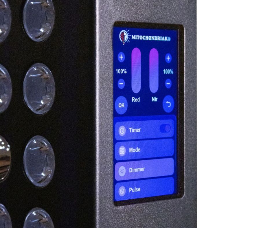
Our Mitochondriak® panels emit wavelengths of 630, 670, 760, 810, 830, 850, and 940 nm. These were chosen to cover both main absorption ranges of CCO: red (heme a, around 620 to 680 nm) and NIR (CuA, around 820 to 850 nm). Wavelengths 630 and 670 nm specifically target the red region of heme a, while 830 and 850 nm specifically target the NIR region of the copper centers.
How does nitric oxide block CCO and how does light release it?
Nitric oxide (NO) is an important signaling gas that serves many functions in the body (vasodilation, blood pressure regulation, immune defense). But in mitochondria, it also has a dark side: it competitively inhibits CCO.
NO binds to the binuclear center of heme a3/CuB at the same site where oxygen normally binds. When NO is bound, oxygen cannot be reduced, electron transport slows, and ATP production drops.
Poyton and Ball (2011) proposed that the release of NO from CCO by the action of red and NIR light is one of the primary mechanisms of photobiomodulation. Photons "displace" NO from the binding site, thereby unblocking CCO and allowing the entire electron transport chain to run at full capacity (Poyton & Ball, 2011).
The released NO then spreads into the surrounding tissue, where it acts as a vasodilator (widening blood vessels and improving microcirculation). So photobiomodulation has a dual effect: it kick-starts the electron transport chain in mitochondria and improves blood flow in the vicinity.
At Mitochondriak®, we also use pulsation (10 Hz), which according to our testing and available data more effectively detaches NO from CCO, because repeated short light pulses prevent its re-binding.
What happens in the cell after light is absorbed by CCO?
When photons of red or NIR light interact with CCO, a cascade of cellular reactions is triggered:
- Increased ATP production – the unblocked CCO transfers electrons faster, the proton gradient strengthens, and ATP synthase works more efficiently
- Increased production of metabolic water (DDW) – more electrons reduce oxygen to water with low deuterium content
- Short-term increase in ROS – a mild increase in reactive oxygen species activates the Nrf2 pathway, the master regulator of the cell's antioxidant defense (hormetic effect)
- Release of NO – vasodilation, improved microcirculation, signaling
- Activation of transcription factors – NF-kB, AP-1, and other factors that regulate gene expression related to regeneration, anti-inflammatory response, and cell proliferation
- Increased mitochondrial membrane potential – an indicator of mitochondrial health
Karu (2008) emphasized that changes in the redox state of CCO lead to "retrograde mitochondrial signaling," meaning signals from mitochondria to the cell nucleus that alter gene expression and influence the overall state of the cell (Karu, 2008).
Why does CCO matter for your health?
CCO is not just a biochemical curiosity. It is the only enzyme in the human body that you can specifically activate with light without any pharmacological intervention. This makes it a bridge between the environment (light) and cellular metabolism.
When CCO functions properly:
- Mitochondria produce enough ATP – you have energy
- High-quality metabolic water with low deuterium is produced
- Oxidative stress is under control
- Cells regenerate and repair efficiently
When CCO is inhibited (by excessive NO, lack of light, toxins, chronic stress):
- ATP production drops – fatigue, poor performance
- ROS accumulate – damage to DNA, proteins, and lipids
- Regeneration slows – slower healing, chronic inflammation
- Overall mitochondrial function deteriorates – the foundation of many chronic diseases
The modern lifestyle (insufficient sunlight, life spent indoors, chronic stress) creates conditions in which CCO is chronically understimulated. Red and NIR light therapy is the most accessible and scientifically supported way to correct this situation. This is precisely what Mitochondriak® panels are designed for, with wavelengths of 630, 670, 760, 810, 830, 850, and 940 nm.
Related glossary terms
- Mitochondria – the cellular organelles where CCO is located
- ATP – the energy whose production CCO directly influences
- Photobiomodulation – red light therapy that acts primarily through CCO
- Circadian rhythm – the biological rhythm influencing the cyclical activity of mitochondria and CCO
- Melatonin – a mitochondrial antioxidant that protects CCO from oxidative damage at night
Stimulate your cytochrome c oxidase with targeted light
Mitochondriak® panels emit wavelengths of 630, 670, 760, 810, 830, 850, and 940 nm, precisely chosen according to the absorption spectrum of CCO. Red light (630, 670, and 760 nm) targets the heme centers, NIR light (810, 830, 850, and 940 nm) targets the copper centers CuA and CuB. Just 10 to 20 minutes per day for more ATP, less inflammation, and better recovery.
- Cytochrome c oxidase (CCO, complex IV) is the final enzyme of the electron transport chain, where oxygen is reduced to water
- It contains four metal centers (CuA, heme a, heme a3, CuB) with copper and iron atoms that absorb red and NIR light
- The action spectrum of CCO according to Karu: peaks at ~620, ~680, ~760, and ~820 to 830 nm
- Mitochondriak® panels emit 630, 670, 760, 810, 830, 850, and 940 nm, specifically covering both absorption ranges of CCO
- Nitric oxide (NO) competitively blocks CCO. Red and NIR light detach NO and CCO starts running again
- Stimulating CCO leads to higher ATP production, metabolic water formation, activation of antioxidant defense, and improved microcirculation
Sources and References
- Hamblin, M. R. (2018). Mechanisms and Mitochondrial Redox Signaling in Photobiomodulation. Photochemistry and Photobiology, 94(2), 199–212. PubMed 29164625
- Karu, T. I. (2005). Absorption measurements of a cell monolayer relevant to the mechanisms of laser phototherapy: reduction or oxidation of cytochrome c oxidase under laser radiation at 632.8 nm. Photomedicine and Laser Surgery, 23(6), 571–577. PubMed 16125966
- Karu, T. I. (2008). Mitochondrial Signaling in Mammalian Cells Activated by Red and Near-IR Radiation. Photochemistry and Photobiology, 84(5), 1091–1099. Wiley doi:10.1111/j.1751-1097.2008.00394.x
- Wong-Riley, M. T. T. et al. (2005). Photobiomodulation Directly Benefits Primary Neurons Functionally Inactivated by Toxins: Role of Cytochrome c Oxidase. Journal of Biological Chemistry, 280(6), 4761–4771. ScienceDirect S0021925820761259
- Poyton, R. O., Ball, K. A. (2011). Therapeutic photobiomodulation: nitric oxide and a novel function of mitochondrial cytochrome c oxidase. Discovery Medicine, 11(57), 154–159. PubMed 21356170
- Ishigami, I. et al. (2023). Structural basis for functional properties of cytochrome c oxidase. PNAS, 120(14), e2216966120. PMC10055264
- Richter, O. M. H., Ludwig, B. (2003). Cytochrome c oxidase – structure, function, and physiology of a redox-driven molecular machine. Reviews of Physiology, Biochemistry and Pharmacology, 147, 47–74. PubMed 12783267